Introduction
Poultry ringworm also known as Favus is a chronic fungal infectious disease commonly associated with several species of dermatophytes belonging to the genera Trichophyton and Microsporum predominantly. The infection occurs when the fungal agent invades the glabrous skin of the birds with their comb being the initial and major nidus of the infection. The infection proceeds as concentrically formed white plaques, which are simultaneously being scaled off and the infection might extend to the feathered regions and sometimes in extreme cases, the condition might become much severe leading up to a point that the fowl succumbs to death.
Etiological Agent (Causative Agent)
The fungi of genera Microsporum and Trichophyton are mainly associated with the infection in fowl and the most reported cases are of
Microsporumgallinae (previously Epidermophyton gallinae ; then Achorion gallinae ; then Lophophyton gallinae ; then Trichophyton gallinae), Microsporum gypseum,Trichophyton megninii and Trichophyton simii
White crest which itself is favus was reported in 7 turkeys out of a flock of 20 and the etiological agent involved was deemed to be a new species which was characteristically intermediate between Trichophyton gallinae and Trichophyton quinckeanum and was thus designated as Trichophyton gallopavum (Metianu et al., 1966).
Epidemiology
Dermatophytosis often occurs due to predisposing factors such as breach in the integrity of the mucosa or skin, exposure to ultraviolet radiations, physico-chemical factors, and presence of underlying disease or uptake of immuno-suppressing medication.
Geographic locations, seasons especially summers and transitioning monsoons and the living conditions affect the prevalence of dermatophytosis. Generally countries with hot and humid climate have are more prone to dermatophytosis and have a higher prevalence. Dermatophytosis is very contagious in wild birds and is often transmitted via direct contact and indirect transmission from environment. (Gochukwu et al., 2022)
Pathogenesis
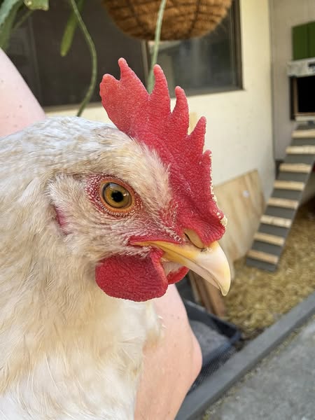
The infection establishes itself at the glabrous skin of comb and wattle of the birds and thereby progresses by the growth of fungal hyphae within the statum corneum of these regions leading to hyperplasia and hyperkeratosis of the epidermal layer. The infection is mostly restricted to the epidermis and is characterized by minimal inflammatory reaction and formation of ‘Favus Cups’ which are depressions around the hair follicles in the feathered areas. The infection often visualized as ‘White Spots’ appear as sprinkled flour and as the infection progresses concentrically, these white spots scale off and give a wrinkled crust appearance to the skin. The appearance of white plaques on comb, face and/or ear lobes is followed by the loss of feathers starting from the caudal base of the comb and continuing down the neck as seen in chicken infected by M.gallinae (Bradley et al., 1993).
In some cases, the respiratory tract is affected with development of nodules and yellow caseous deposits.
Histopathological Lesions and Clinical Signs
The infection manifests itself by invasion of the hairless areas on the body of fowl and there are various histopathological lesions observed upon microscopic examination which often include:
•Hyperkeratosis of skin epithelium with invasion of stratum corneum by fungal mycelium
•Acanthosis
•Acantholysis
•Hydropic degeneration of the cells in stratum spinosum
•Underlying dermis is often infiltrated by mononuclear cell and has lymphoid foci
•The infection is generally limited to the epidermis and gives appearance of sprinkled flour due to the serocellular crusts with primary component of heterophils mixed with fungal mycelia
• Multifocal suppurative folliculitis
• Chronic lymphoplasmacytic and histiocytic perifolliculitis
Diagnosis
The diagnostic techniques for these infectious agents include the fungal culture examination on Sabouraud Dextrose Agar (SDA) or Selective Dermatophyte Media followed by staining with Periodic acid-Schiff stain (PAS stain) or with Gomori’s methanamine silver stain. The sample is derived from the externally visible lesions and swabs from deeper layers of fungal penetration. The morphology of hyphae (septate) and pink stained conidia (macroconidia and microconidia) are the observed upon microscopic examination.
Prevention
The infection is of fungal origin and the fungi grow at optimum and often accelerated rates in a relatively warm and moist surfaces, thus if these conditions are not available, then their growth can be retarded and as the infection is likely to rescind on its own, not much intervention is required.
The disease is known to spread from the infected bird to the healthy ones in the flock, thus mass infection can be avoided by simple steps of prevention. Favus can also be prevented by managemental practice of ‘dubbing’ which is the removal of part of comb in birds generally done via a pair of scissors, dubbing is best performed in day old to one week old chicks, in order to control the bleeding.
Control
The infection is less observed in the intensive poultry but the backyard birds and the fowl of free range flocks are much more susceptible hosts for the fungal infection. The condition affects individual birds from within a flock but is transmitted to the healthy flock members by gradual physical contact. Contamination of the housing material is an important factor in spread of the infection. Thus, the segregation and isolation of the infected individuals and disinfection are crucial control methods.
The infection is often reported to rescind without veterinary intervention and thus is said to be self limiting in nature and is reported to heal within 10 weeks.
Treatment
The infection is often self controlled but in some cases might need medicinal treatment. Generally, topical antifungals like miconazole, enilconazole, cleaning the lesions with povidone iodine and strategies for general supportive treatment are advised. Various tested treatments include: removal of crusts from the skin and treating the infected area with topical antifungal compounds and dressing the area. Azole compounds have proved to be effective in the treatment of avian dermatophytosis. Their mechanism of action is by inhibiting the conversion of lanosterol to ergosterol, which is an integral component of the fungal cell membrane. The drugs of this class act upon lanosterol 14-alpha-demethylase (CYP51) enzyme in the fungal cell membrane which is responsible for the conversion of lanosterol to ergosterol and as a result, the cell membrane becomes more permeable and the cells lyse and die.
Treatment of the infected area has been successful by topical application of miconazole (Droual et al., 1991), 2% miconazolenitrate (Bradley et al., 1995), tincture of iodine (Mustaffa-Babjee and Oommen, 1970). An ointment made of liquefied vaseline and 5% formaldehyde when rubbed over the lesion resulted in successful treatment (Beach and Hairpin, 1918; Chute, 1972). Single application of Quaternary Ammonium Compounds have proved to be effective in treating favus in poultry. Clove essential oil ointment (3% w/w) was found to be as effective and non-inferior to a 2% w/w ointment of ketokonazole in M.gallinae infected chicken (Sucheeva et al., 2021). 90% birds were found to be culture negative and completely recovered on day 35 after initial treatment.
Conclusion
Favus or avian ringworm is hardly relevant in commercial poultry. However, due to the exposure of backyard poultry to other avians in recent years, it’s an important diagnosis which should never be overlooked. Periodic disinfection along with maintaining hygiene and sanitation of the farm will effectively prevent and control the disease.
Ishan Garg*, Amitav Bhattacharyya and Pankaj Kumar Shukla
College of Veterinary Science and Animal Husbandry, DUVASU, Mathura, India
*E mail: ishan25g@gmail.com


