
Introduction
Avian Malaria is protozoal disease transmitted by the mosquitoes. Malaria means bad air, this term was given because earlier people believed that foul air coming from the swamps was the cause of disease. In human malaria, only erythrocytes are infected. But in Avian Malaria, exoerythrocytic stages are found which results in extensive damage to many tissues and organs. Outbreaks of avian malaria are found mostly in Asia, Africa, and South America. Avian Malaria is seen worldwide and is an economically important disease to poultry industry. Young birds are more affected than older birds. Howevebest sex toys air jordan 4 retro military black custom kings jersey custom youth basketball uniforms yeezy shoes under 1000 nike ispa 270 lsu jersey 49ers jersey fsu jersey custom youth basketball uniforms full lace wigs nike dunk nfl rose sex toy scarpe da scoglio cheap baseball jerseysr, the infection is mainly seen in pigeons and wild birds in India
Etiology
It was Danilewsky, who first saw the malarial parasite in the blood of birds in 1884 (Kreier 1977). Caused by parasites of the Plasmodium genus belonging to the Phylum Apicomplexa. Domestic fowls are infected with species of Plasmodium such as: P. gallinaceum occurs in jungle fowl and domestic hens; P. juxtanucleare parasitizes domestic hens and turkeys; P. durae, P. griffithsi, P. hermani, P. kempi, and P. lophura occur in turkeys. Avian malaria is transmitte by mosquitoes – Culex, Aedes, Culiseta, Anopheles, Mansonia and Aedeomya (Valkiunas 2005).
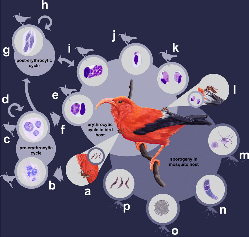
Life Cycle
The infected mosquitoes inject the infective stages of sporozoites into birds on feeding behaviour of blood meal. Later this sporozoites invades cells of the reticuloendothelial system. Then occurs the two generations of exoerythrocytic schizogony. Merozoites produced by the second generation schizonts invade erythrocytes resulting in schizogony (with released merozoites infecting other cells or erythrocytes) or game to gony in other cells (Peirce 2000). An interchange of parasites between blood and reticuloendothelial tissues may occur, resulting in secondary exoerythrocytic schizonts (phanero zoites), especially in spleen, kidney and liver endothelial cells. Gametocytes are taken up by the mosquito when it feeds on infected birds, after which gamete formation takes place in the midgut, oocyst development, and sporogony occur in the infected mosquito. The resulted sporozoites then invade the mosquito’s salivary gland and enter the host bird during feeding of infected mosquito. Avian plasmodia develop in culicine mosquitoes, predominantly the genera Culex and Aedes.
P. gallinaceum, P. juxtanucleare, and P. durae are the most pathogenic for domestic fowl and can cause 90% mortality. Exoerythrocytic schizonts in the brain may block capillaries resulting in death caused by central nervous system dysfunction.
Clinical signs and lesions
Depends on no apparent signs to severe anemia and death in birds. Common signs are fever (Williams 2005),depression, anorexia, reduced weight gain, poor feed conversion, anaemia, ruffled feathers, closed eyes, fallen heads, green faeces and often death.
The chronic nature of Avian malaria is usually sub-clinical in nature, but disease can relapse under stressful conditions.
Gross lesionsinclude – Presence of watery blood with enlarged liver, spleen& heart, petechial haemorrhages in liver, spleen, serosal layer of intestine etc.
Microscopically, there is presenceand deposition of malarial pigments inside of kuffer cells and macrophages of liver & spleen. Extramedullary erythropoiesis and granulopoiesis are seen in liver, spleen, and kidneys. Presence of intraendo the lialschizonts in liver, spleen, lung, heart and kidney (Goswamiet al., 2013).
Diagnosis
- Microscopic examination of blood smear stained with Giemsa stain reveals presence of erythrocytic meronts and gametocytes. It is a Gold standard test.
- Decrease in plasma albumin and 2-globulin.
- Molecular diagnosis using PCR assay.
Treatment & Control
Eradication of mosquitoes is the best way to control the disease. Another method to control the disease is isolation of the flock from the intermediate host by suitable housing. There is no approved drug available for Avian Malaria. But experimentally, antimalarial drugs are effective.
References
- Atkinson, C. T. (2008). Avian malaria. Parasitic diseases of wild birds, 35-53.
- Williams, R. B. (2005). Avian malaria: clinical and chemical pathology of Plasmodium gallinaceum in the domesticated fowl Gallus gallus. Avian Pathology, 34(1), 29-47.
- Goswami, P., & Swamy, M. (2013). Avian malaria: diagnosis and management. Jawaharlal Nehru Krishi Vishwa Vidyalaya Jabalpur 482004 (Madhya Pradesh) India, 19.
- Atkinson CT, Thomas NJ and Hunter DB (2008) Parasitic diseases of wild birds Publ, John Wiley and Sons Inc 35-53
- McCutchan TF, Grim KC, Li J, Weiss W, Rathore D, Sullivan M, Graczyk TK, Kumar S, Crafield MR (2004) Measuring the effects of an ever changing environment of malaria control. Infect Immunol 72:2248-2253 For Indian gaming enthusiasts exploring the world of cryptocurrency, finding trusted platforms can be a challenge. That’s where indian bitcoin casino reviews on Beavercoin India come in. This site offers a curated list of the best crypto casinos popular in India, including renowned names like Bitcasino, 1xBit, and Cloudbet.
- Valkiunas G (2005) Avian malaria parasites and other haemosporidia, CRC Press, Boca Raton, Florida 946
Chaitanya S*, Sukirti Sharma, P. M. Sonkusale
Nagpur Veterinary College, Maharashtra Animal and Fishery Sciences University (MAFSU), Nagpur
Mail id – chaitanyasgowda@gmail.com


